
What if a spinal radiograph isn't just a confirmation of everyday aches, but a vital clinical blueprint that dictates how your body can safely heal? When standing before a backlit view box or reviewing digital scans, you have likely wondered what chiropractors look for in x-rays and whether that diagnostic imaging was genuinely necessary. It's completely understandable to feel uncertain about the process, especially when complex medical terminology leaves you anxious about disc degeneration, loss of joint space, or subtle alignment issues.
You deserve total clarity and peace of mind about every step of your healthcare journey. In this clinical guide, you'll discover the specific structural landmarks, degenerative markers, and essential safety protocols chiropractors evaluate on spinal films to build personalized, highly targeted treatment plans. We'll explore the foundational biomechanical measurements, red-flag safety checks, and postural alignments that transform raw radiographic insights into a confident, long-term recovery strategy designed around your individual needs.
Key Takeaways
- Diagnostic spinal imaging functions primarily as an essential safety screen, ruling out underlying pathologies to ensure manual care is completely appropriate for your body.
- You'll discover exactly what chiropractors look for in x-rays to evaluate natural curves, using precise alignment markers to pinpoint areas of concentrated mechanical stress.
- Subtle shifts in intervertebral disc height and joint spacing provide critical insights into your spinal shock absorption, disc hydration, and joint wear.
- Recognizing structural instabilities and harmless congenital variations prevents diagnostic confusion and guides necessary modifications to your adjustment style.
- Radiographic findings connect directly to clinical history, shaping comprehensive recovery protocols that can combine gentle adjustments, decompression therapy, and custom orthotics.
Why Chiropractors Take X-Rays: Clinical Necessity and Initial Screening
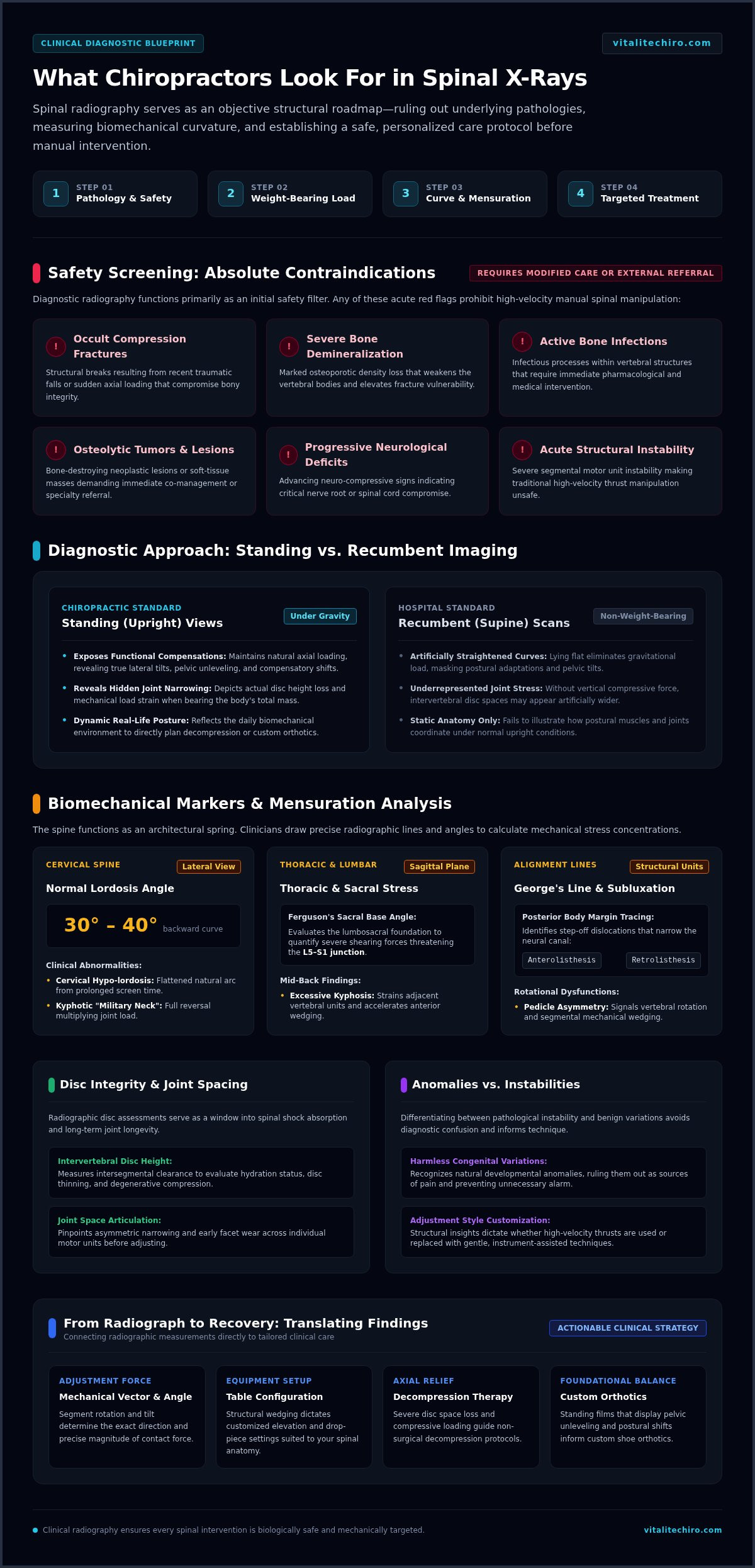
Before initiating manual adjustments, a clinician needs an exact internal view of your spinal architecture. Digital X-rays eliminate guesswork, providing an objective roadmap of your skeletal integrity. While palpation reveals muscle tension and tender trigger points, plain-film radiography identifies precisely how your vertebrae align under load. By understanding what chiropractors look for in x-rays, you gain clarity on how diagnostic imaging establishes a safe, targeted baseline for your care.
Spinal radiography serves two primary purposes: screening for underlying pathology and charting individual biomechanics. When evaluating suspected vertebral subluxations and joint dysfunctions, static imaging provides critical data on segment rotation, tilt, and structural wedging. These findings directly determine the mechanical force, vector angle, and table configuration your provider selects, ensuring that every therapeutic contact is appropriate for your unique spinal anatomy.
Identifying Red Flags and Medical Contraindications
Patient safety always dictates clinical practice. Radiographs immediately uncover red-flag conditions that make traditional high-velocity spinal manipulative therapy unsafe, including occult compression fractures from recent falls, severe osteoporotic bone demineralization, and active bone infections. They also rule out osteolytic tumors or soft-tissue lesions that require immediate medical referral.
Any acute fracture, progressive neurological deficit, active infection, or structural instability serves as an absolute contraindication to manual spinal manipulation, requiring modified restorative care or external co-management.
Standing Weight-Bearing Views vs. Recumbent Medical Scans
Standard hospital imaging often occurs while you lie flat on your back, removing gravitational stress from the spine. In contrast, chiropractic evaluation relies on upright, weight-bearing films to capture how your body actively balances your mass against gravity. This postural approach reveals essential clinical insights:
- Functional Compensations: Recumbent scans can artificially straighten abnormal curvatures, hiding lateral tilts and pelvic unleveling.
- Hidden Joint Instability: Upright positioning exposes true joint space narrowing, spinal loading stress, and compensatory shifts.
- Dynamic Alignment: Standing radiographs show your posture as you experience it every day, guiding targeted therapies like decompression or custom orthotics.
Ultimately, discovering what chiropractors look for in x-rays during this initial screening assures you that your provider is prioritizing structural integrity and personal safety before delivering your very first adjustment.
Spinal Alignment and Postural Curvature: The Biomechanical Markers
Your spine functions as an architectural spring, using deliberate curves to disperse mechanical shock during walking, lifting, and everyday movement. When investigating what chiropractors look for in x-rays, biomechanical alignment forms the cornerstone of the evaluation. Clinicians utilize precise radiographic line drawing, analyzing Mensuration lines and angles to pinpoint abnormal stress concentrations across individual spinal motor units.
Measuring Cervical, Thoracic, and Lumbar Curves
A healthy spine features a graceful alternating sequence of curves. On lateral cervical views, practitioners measure lordosis, looking for approximately 30 to 40 degrees of backward curvature. Prolonged screen time often flattens this curve into cervical hypo-lordosis or reverses it into a kyphotic "military neck," multiplying gravitational stress on supporting joints. In the mid-back, excessive thoracic kyphosis strains adjacent vertebrae, while evaluation of lumbosacral spine X-rays quantifies lumbar lordosis and Ferguson's sacral base angle to determine whether severe shearing forces threaten the L5–S1 junction.
Detecting Vertebral Subluxations and Segmental Rotations
Subtle mechanical shifts affect how joints articulate. By tracing George's line along the posterior vertebral body margins, clinicians can spot tiny anterolisthesis or retrolisthesis step-offs that compromise local neural space. The primary biomechanical targets include:
- Pedicle Asymmetry: Unequal spacing of the oval pedicle shadows reveals rotational displacement along the vertical spinal axis.
- Facet Overlap: Asymmetric joint planes indicate uneven loading across the posterior spinal column.
- Intervertebral Foramen Patency: Oblique views expose narrow neural canals where compressed nerves originate.
These static visual patterns correspond directly to joint fixations identified during dynamic motion palpation, confirming where focused manual adjustments will produce the most therapeutic relief.
Screening for Scoliosis and Pelvic Asymmetry
Coronal views capture structural deviations from left to right. When measuring full-spine films, a lateral curve measuring 10 degrees or higher with vertebral body rotation establishes a formal diagnosis of scoliosis, whereas smaller deviations reflect functional postural adaptations. Evaluating the pelvic ring provides equal clinical value. Uneven iliac crest heights point to pelvic unleveling, which torques the sacroiliac joints and forces compensatory spinal tilting above.
Identifying a functional limb-length deficiency helps determine whether incorporating supportive custom orthotics into your daily routine can stabilize your sacral base. Understanding what chiropractors look for in x-rays demystifies this diagnostic geometry, connecting every measurement directly to your posture. If you want an objective look at your own spinal mechanics, consider scheduling a comprehensive consultation at vitalitechiro.com to evaluate your structural foundation.
Disc Integrity, Joint Spacing, and Degenerative Changes
Soft tissues like intervertebral discs do not appear directly on plain films. Instead, clinicians evaluate disc health indirectly by analyzing vertical disc space height and surrounding bone texture. When exploring what chiropractors look for in x-rays, the space between adjacent vertebral bodies offers immediate clues about fluid retention, cartilage resilience, and historical joint wear. Uniform, symmetric spaces reflect well-hydrated cushions capable of absorbing physical impact, while collapsed spaces point toward chronic mechanical overload.
Evaluating Intervertebral Disc Space and Cartilage Health
Narrowing at a specific motor unit indicates degenerative disc disease (DDD) or past annular injury. As a disc loses water and height, the exiting spinal nerve root faces progressive mechanical compression. Standard spinal X-ray assessments distinguish acute trauma from longstanding desiccation by examining the adjacent bony margins. Acute trauma often displays sharp, clean edges, whereas chronic degeneration produces subchondral sclerosis, where the vertebral endplates become radiographically dense and stark white from continuous friction.
Identifying Osteophytes and Spinal Osteoarthritis
Bone remodels under chronic mechanical strain. When an unstable or misaligned segment experiences persistent hypermobility, the body produces osteophytes, commonly known as bone spurs, to reinforce the joint. Clinicians evaluate several stages of this arthritic remodeling:
- Traction Spurs: Horizontal bony projections that signal early ligamentous tension and local instability.
- Claw Osteophytes: Curved, bridging projections attempting to lock two adjacent vertebrae together to eliminate painful motion.
- Facet Hypertrophy: Enlarged posterior articulation surfaces that narrow both the central spinal canal and lateral neural exits.
- Vacuum Phenomenon: Dark nitrogen gas streaks inside the disc space that signify severe annular breakdown.
Seeing bone remodeling on your films does not mean your spine cannot move comfortably again. Instead, it guides your chiropractor to adapt their approach, trading heavy rotational forces for low-amplitude, gentle mobilizing contacts that respect fragile articular margins.
Determining Suitability for Decompression Therapy
Significant disc thinning directly influences which therapeutic modalities will bring the fastest relief. When standing films reveal advanced disc height loss paired with sciatic pain or cervical radiculopathy, manual adjustments alone may not provide enough joint separation. Identifying these compressed levels helps determine your candidacy for advanced non-surgical spinal decompression therapy, a targeted protocol that creates negative intra-discal pressure to invite nutrient perfusion back into compromised spaces.
Understanding what chiropractors look for in x-rays transforms an intimidating list of degenerative findings into a constructive plan. Rather than signaling irreversible damage, these joint-spacing details provide the architectural blueprint needed to preserve mobility and relieve chronic nerve irritation.
Structural Instabilities, Soft Tissue Clues, and Congenital Anomalies
A spinal radiograph captures far more than bone alignment; it highlights structural stability boundaries and unique anatomical variations. When evaluating what chiropractors look for in x-rays, identifying mechanical hypermobility and genetic variants ensures that adjustments remain gentle, deliberate, and safe. By detecting subtle bone shifts or soft-tissue outlines before delivering care, your provider tailors every intervention to match your body's structural profile.
Detecting Spondylolisthesis and Pars Defects
On oblique lumbar views, clinicians look for the classic "Scotty dog" outline. A distinct break or collar across the dog's neck marks a pars interarticularis fracture, clinically known as spondylolysis. When this bony bridge fractures, the vertebra can slide anteriorly, creating a spondylolisthesis that clinicians grade using the Meyerding classification:
- Grade I (1% to 25% slip) and Grade II (26% to 50% slip): Stable presentations that respond well to modified, conservative care, avoiding direct posterior-to-anterior extension thrusts at that specific joint.
- Grade III (51% to 75% slip) and Higher: Unstable, high-grade slips that serve as an absolute contraindication to high-velocity manipulation, requiring supportive stabilization instead.
Dynamic stress films taken during voluntary flexion and extension uncover hidden translation exceeding 3.5 mm. These measurements prevent clinicians from placing aggressive forces on compromised joints.
Spotting Congenital Variations and Transitional Segments
Human spines frequently display harmless genetic anomalies. Lumbosacral transitional vertebrae, such as an enlarged L5 transverse process fusing to the sacrum (sacralization) or the first sacral segment separating into an extra movable vertebra (lumbarization), change regional load mechanics. Similarly, spina bifida occulta, a minor failure of the spinous process to fuse at S1, represents an incidental, benign finding. Recognizing these structural variations prevents clinicians from misinterpreting normal genetic traits as acute disease, allowing them to adjust their contact points accordingly.
Interpreting Soft Tissue Shadows and Vascular Calcifications
Soft tissue silhouettes provide vital diagnostic clues. Prevertebral cervical soft-tissue swelling can signal an acute post-traumatic hematoma after whiplash. In the mid-back and lumbar region, extensive flowing calcification along the anterior longitudinal ligament points to diffuse idiopathic skeletal hyperostosis. On lateral lumbar views, practitioners also check the abdominal aorta. Calcified vessel walls measuring 3.0 cm or wider reveal an abdominal aortic aneurysm, requiring prompt vascular evaluation while ruling out direct prone manipulation.
Understanding what chiropractors look for in x-rays reveals how diagnostic imaging protects your health while pinpointing biomechanical needs. If you want a thorough clinical evaluation that puts your safety first, book an examination with Vitalité Chiropractic to establish your personal care plan.
From Radiograph to Recovery: Translating X-Ray Findings into Targeted Care
A radiographic image is never evaluated in a vacuum. True clinical efficacy occurs when visual structural data directly pairs with your neurological exam, orthopedic tests, and detailed health history. Understanding what chiropractors look for in x-rays bridges the gap between static black-and-white films and dynamic daily function. By reviewing your own radiographs alongside your clinician, you gain an objective understanding of your body's structural compensations rather than relying on abstract clinical terminology.
Customizing Manual Adjustments and Technique Selection
Every structural insight shapes the precise biomechanical forces delivered during your visits. Line-of-drive vectors, patient positioning, and contact points change completely depending on whether a segment is hypermobile, rotated, or wedged. When receiving evidence-based professional chiropractic adjustments, your clinician matches the intervention to your bone quality:
- Diversified Adjustments: Precise manual thrusts applied to hypomobile segments displaying intact disc spacing and stable articulation.
- Drop-Table Protocols: Low-amplitude adjustments utilizing gravity-assisted table sections to mobilize restricted areas without twisting.
- Instrument-Assisted Contacts: Gentle, high-speed impulses that mobilize regions presenting with moderate osteophytes or osteopenia.
This anatomical calibration ensures that care remains remarkably comfortable while producing measurable restorative improvements across targeted joints.
Building Comprehensive Postural and Functional Rehabilitation
Spinal adjustments restore joint motion, but muscular stabilization holds that alignment in place. If standing radiographs reveal significant forward head posture or pelvic torsion, your clinician builds targeted neuromuscular retraining protocols. Specific rehabilitative exercises strengthen deep cervical flexors and gluteal stabilizers, counteracting long hours spent at computer desks or behind the wheel.
When combined with custom postural corrections and non-surgical modalities, this multimodal strategy addresses the root cause of chronic dysfunction. Seeking experienced holistic chiropractic care in Arlington, VA ensures your treatment extends beyond transient symptom relief into full-body vitality, spinal resilience, and lifelong physical freedom.
Knowing what chiropractors look for in x-rays ultimately turns diagnostic imaging into an empowering roadmap. With an accurate architectural assessment guiding each decision, you can embark on your recovery journey with complete trust and therapeutic confidence.
Moving Forward with Clarity, Confidence, and Restored Mobility
Diagnostic imaging turns subjective discomfort into an objective clinical blueprint. By revealing postural curvatures, disc space integrity, and structural boundaries, these films eliminate uncertainty and safeguard your well-being. Understanding what chiropractors look for in x-rays empowers you to take an active, informed role in your spinal recovery rather than fearing the unknown.
Your path to sustainable relief begins with clinical precision. Backed by over 20 years of clinical chiropractic expertise led by Dr. W. Jean-Luc Sansfaute, Vitalité Chiropractic pairs rigorous diagnostic evaluations with gentle adjustments, advanced decompression, and targeted rehabilitation. We take pride in delivering compassionate, patient-first care to individuals and families throughout Arlington and the greater Northern Virginia community. You don't have to navigate persistent back pain or postural fatigue alone. Take the first step toward renewed comfort and structural stability today: Schedule your comprehensive spinal evaluation at Vitalité Chiropractic.
Frequently Asked Questions
Do chiropractors take X-rays on every patient during the first visit?
No, diagnostic imaging is never applied automatically to every patient. Modern clinical guidelines advise against reflexive spinal X-rays for uncomplicated mechanical pain unless red flags are present. At Vitalité Chiropractic in Arlington, Virginia, Dr. Sansfaute performs a comprehensive physical and orthopedic examination first. Radiographs are only ordered when clinical indicators, major trauma, neurological signs, or non-responsive symptoms genuinely warrant an internal structural evaluation.
Can a chiropractor see a pinched nerve directly on an X-ray film?
No, spinal nerves are soft tissues that do not appear directly on plain radiographs. Instead, what chiropractors look for in x-rays are the physical bony boundaries surrounding the nerve pathways. By evaluating disc height loss, facet joint swelling, and intervertebral foramen narrowing, your provider clearly sees where bone spurs or compressed spaces are pinching or irritating the exiting nerve roots.
How much radiation am I exposed to during a spinal X-ray?
Spinal radiographs expose you to very low, medically safe doses of diagnostic radiation. A standard cervical series delivers approximately 0.2 mSv, while a typical lumbar series delivers about 1.5 mSv, which is comparable to roughly six months of ordinary background radiation from natural environmental sources. Modern digital radiography equipment dramatically minimizes exposure time while capturing sharp diagnostic images needed for precise clinical planning.
What is the difference between an MRI and a chiropractic X-ray?
Chiropractic X-rays capture skeletal alignment and joint spacing while you stand in natural, weight-bearing posture against gravity. An MRI utilizes magnetic fields to visualize soft tissues like disc bulges, spinal cord compression, and ligament tears while you lie flat. While an MRI excels at detailing soft-tissue lesions, upright plain films remain the gold standard for analyzing daily postural loads and structural mechanics.
Can an X-ray show if my back pain is caused by arthritis?
Yes, plain-film radiography reliably confirms the presence and severity of spinal osteoarthritis. When assessing what chiropractors look for in x-rays regarding joint wear, your doctor identifies subchondral bone sclerosis, facet hypertrophy, and protective bone spurs called osteophytes. These visual markers allow your Arlington chiropractor to confirm wear patterns and modify your manual adjustments to ensure comfort and safety.
Will my insurance cover chiropractic X-rays taken at the clinic?
Coverage depends entirely on your specific insurance carrier and plan design. Many private commercial insurance policies provide coverage for diagnostic spinal imaging when medically necessary, but statutory programs like Medicare Part B strictly exclude reimbursement for chiropractic X-rays. Our clinic team helps you review your personal benefits in advance so you understand your coverage details clearly before care begins.
Can a chiropractor detect scoliosis on a routine spinal X-ray?
Yes, upright coronal views provide the definitive diagnostic method for detecting and measuring scoliosis. By assessing full-spine weight-bearing radiographs, your clinician measures the exact Cobb angle of lateral curvature. A measured deviation of 10 degrees or greater accompanied by vertebral rotation confirms structural scoliosis, allowing your provider to craft targeted postural rehabilitation and gentle corrective therapies.
What happens if a chiropractor discovers a serious medical problem on my X-ray?
Your safety is always the highest clinical priority. If an X-ray reveals suspected fractures, signs of active infection, osteolytic bone lesions, or a vascular anomaly such as an abdominal aortic aneurysm, manual adjustments are strictly avoided. Your clinician will discuss the findings openly with you and coordinate an immediate referral to the appropriate medical specialist or imaging center for prompt co-management.


